
|
|
How to determine Fermi vectors by angle resolved photoemission? |
![]()
|
A new method for the determination of Fermi vectors from photoemission
spectra is presented. It produces Fermi surfaces with high accuracy and
reliability even for narrow band systems. A wide variety of physical phenomena of materials rely on details of the topology of the Fermi surface. However, traditional experimental determinations are restricted to bulk materials. More complex cases, such as superlattices, heterostructures or even clean surfaces can hardly be accessed. In such situations, angle-resolved photoemission spectroscopy (ARPES) has recently been extensively applied to gain insight into the topology of Fermi surfaces.
Such a method is presented here as a result of a strong collaboration between experiment and theory. The new method had to be simple and stable for the necessities of the measurement and reliable from a theoretical point of view. Employing high resolution photoemission spectroscopy we show how surface-parallel components of the Fermi vectors can be determined with high accuracy. Intensity modifications due to the photoemission process are explicitly eliminated by comparing photoemission spectra taken at different temperatures. If from one of the other methods a rough estimate is known, the Fermi vector can accurately be fixed by subtracting the photoemission intensities for two temperatures. If taken at the Fermi level for different wave vectors in the neighborhood of the estimate, the vanishing of the difference determines the Fermi vectors. This is valid for a variety of spectral functions, including Lorentzians, Gaussians, Voigt profiles, the Luttinger model, a refined Luttinger profile, two dimensional and marginal Fermi liquids.
|
 |
 |
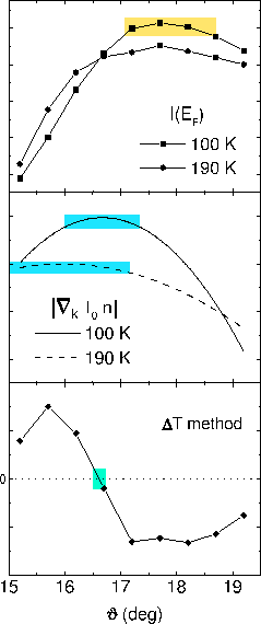
| Fig. 1: Angle resolved photoemission spectra associated with the
Ti 3d-band of 1T-TiTe2 in the Gamma-M
direction were taken at 100 K
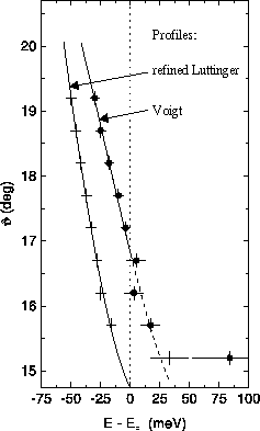
and 190 K. Left: (top ) Intensities at the Fermi level (maximum-intensity method). Yellow bar marks the uncertainties for the parallel part of the Fermi vector (95% of the maximum value). (middle) Smoothed k-derivatives of the intensities integrated over the whole spectra (maximum-gradient method). Blue bars mark the uncertainties as above. (bottom) Difference of the two intensities at the Fermi level. The intersection with zero marks the Fermi vector. The green bar shows the uncertainty of 0.1° which emerges from the error of the Fermi energy (1 meV). Right: kF determination from extrapolation of fitted peak positions using two different profiles.
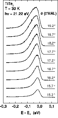
For an application of the new method we study normalized photoemission spectra
of the layered material 1T-TiTe2, taken
at 100 K and 190 K. The high energy and angle resolved spectra
along the Gamma-M direction contain the peaks emerging from the
Ti 3d-band, which is well separated from contributions of other bands.
Intensities at the Fermi level
are depicted in Fig. 1, top left. According to the new method
the Fermi vector is given by the intersection of the
curves, which can clearly be identified at a
value of 16.6°, see the difference of the spectra at the bottom of the left
panel.
For comparison we also show the values
obtained from the maximum intensity (top) and maximum gradient (middle)
methods as described above. The maximum intensity at
the Fermi level can be observed on a relatively broad peak
at around 17.8°, showing a systematic erroneous shift
towards occupied states. The derivative of the energy
integrated intensities are depicted in
the middle of the left panel. Since the gradient of the
integrated intensity is only marginally
changing in the regime of interest a
rather broad maximum is observed making a detailed
quantitative analysis
difficult. In the right panel estimates from the peak-tracing method for
two assumed profiles are shown. With a Voigt profile the Fermi vector
appears at 16.7° and with a refined Luttinger profile at 14.75°.
See also:
Acknowledgment:
This research is supported by the BMBF, FR Germany
(project No. 05 SB8 FKB, 05 SE8 FKA and
05 SB8 FKA).
|
 Fermi vectors have been extracted from experimental ARPES data like those
shown on the left, employing three kinds of criteria,
Fermi vectors have been extracted from experimental ARPES data like those
shown on the left, employing three kinds of criteria,